 首页
首页 400-620-6333
PICO ECL,超敏发光液
Pico ECL底物可为使用辣根过氧化物酶(HRP)偶联物的免疫印迹实验提供明亮的信号,该ECL底物能够兼容各种膜、封闭液和宽范围抗体稀释液,以出色性能、通用性和高性价比,满足用户的免疫印迹应用需求。
有货
| 英文名称 | Pico ECL |
|---|---|
| 储存温度 | 2-8°C储存,避光 |
| 运输条件 | 冰袋运输 |
| 产品介绍 |
包装规格:100ml: 一抗50ml+二抗50ml;500ml:一抗250ml+二抗250ml Pico ECL底物可为使用辣根过氧化物酶(HRP)偶联物的免疫印迹实验提供明亮的信号,该ECL底物能够兼容各种膜、封闭液和宽范围抗体稀释液,以出色性能、通用性和高性价比,满足用户的免疫印迹应用需求。 ?Pico ECL底物的特点: ? Pico ECL —用于辣根过氧化物酶(HRP)的增强型化学发光底物 ? 低皮克级灵敏度—检测硝化纤维素膜或PVDF膜上低皮克级的蛋白条带 ? 长信号持续时间— 在条件优化情况下,经底物孵育的印迹条带能够持续输出6至8小时的可检测光信号 ? 稳定试剂— 工作液在24小时内保持稳定;试剂盒在室温下可稳定放置长达1年 ? 价格经济— 针对稀释的抗体浓度条件进行了优化: ? 0.2至 1.0 μg/mL一抗(以1 μg/mL储存液稀释1:1,000至1:5000倍) ? 10至 50 ng/mL二抗(以1 μg/mL储存液稀释1:20,000至1:100000倍) ? 重要提示 1为获得最佳效果,必须优化该系统的全部组分,包括样品量、一抗和二抗浓度以及膜和封闭试剂的类型。 2?使用该产品比使用沉淀比色HRP底物检测所需的抗体浓度低。为优化抗体浓度,请进行一次系统的点印迹分析。 3没有一种封闭试剂对所有系统而言都是最佳的,所以为每一个免疫印迹检测系统找到最合适的封闭缓冲液非常必需。封闭试剂有可能与抗体产生交叉反应,导致出现非特异性信号。封闭缓冲液同时也会影响系统的灵敏性。当从一种底物转换为另一种底物时,有时会出现信号衰减或背景增加的现象,原因可能是封闭缓冲液不适合新的检测系统。 4?使用亲和素/生物素检测系统时,避免使用牛奶作为封闭试剂,因为牛奶中含有不定量的内源性生物素,会导致高背景信号。 5??保证洗涤缓冲液、封闭缓冲液、抗体溶液和底物工作液的使用体积,以确保在整个实验过程中印迹膜完全被液体覆盖,避免膜变干。增大封闭缓冲液及洗涤缓冲液的使用量可以降低非特异性的信号 6??为获得最佳效果,在孵育步骤请使用摇床。 7??将Tween20(终浓度0.05-0.1%)加入封闭缓冲液和稀释的抗体溶液,以降低非特异信号。使用高品质的产品,如去污剂。它保存在安瓿中,过氧化物和其他杂质含量很低。 8??不要使用叠氮钠作为缓冲液的防腐剂。叠氮钠是HRP的抑制物 9??避免手与膜直接接触,实验过程应戴手套或使用干净的镊子 10??所有设备必须清洁且不沾染外来物质。金属器械(如剪刀)不得具有可见的锈迹。锈迹可能导致斑点形成和高背景。 11??底物工作液在室温下可稳定8小时。日光或任何其他强光下可能损害底物,为获得最佳结果,将底物工作液保存在琥珀色瓶中,并避免长期暴漏在任何强光下,短时间暴漏于实验室常规照明不会损害该工作液 ? 操作概述 ??注:优化抗原和抗体的浓度。必须使用建议的抗体稀释度,以保证阳性结果。有关建议的稀释度范围请参考其他所需材料。 1)??将一抗浓度稀释到0.2~1ug/ml 2)?将二抗浓度稀释到10~50ng/mL 3)?将两种底物组份按1:1比例混合,制备底物工作液。 注:暴漏于日光或任何其他强光可能损害工作液,为获得最佳结果,将此工作液保存在琥珀色瓶中,并避免长期暴漏于任何强光。短时间暴漏于实验室常规照明不会损害该工作液。 4)?将印迹膜在West Pico ECL底物工作液中孵育5分钟。 5)?吸出多余试剂。用清洁的塑料膜盖住该印迹膜。 6)??使印迹膜在X光胶片上曝光。 ? 其他所需材料 l??已完成转印的印迹膜:用合适的电泳法分离蛋白质,并将这些蛋白质转移到硝酸纤维素膜上。 l??稀释缓冲液:使用Tris或磷酸盐缓冲液。 l??洗涤缓冲液:将5mL 10%的Tween-20加入1000mL稀释缓冲液(Tween-20的终浓度将0.05%)。 l??封闭试剂:将0.5mL10%的Tween-20加入100mL的封闭缓冲液,选择一种与稀释缓冲液具有相同基本组分的封闭缓冲液。 l??一抗: 选择一种目标蛋白质特异性抗体。使用稀释缓冲液制备该抗体的1ug/ml储备液。使用封闭试剂将抗体从储备液稀释成抗体工作液。稀释度介于1:1000和1:5000之间或抗体工作液浓度为0.2~1ug/ml.最佳稀释度取决于一抗和膜上的抗原量。 l??HRP标记的二抗:选择一种与二抗特异性结合的HRP标记二抗,使用稀释缓冲液制备该抗体的1ug/ml储备液。使用封闭试剂将抗体从储备液稀释成抗体工作液。稀释度介于1:20000和1:100000之间或抗体工作液浓度为10~50ng/ml。该浓度范围在使用链亲和素-HRP时也适用。二抗的最佳稀释度取决于HRP标记二抗和膜上的抗原量。 l??用于处理放射显影胶片的胶片暗盒、显影和定影试剂 l??用于孵育的旋转摇床。 ? 蛋白印迹法详细操作步骤 1)?将印记膜从蛋白转印设备中取出,加入合适的封闭液在温室下孵育20-60分钟,同时振荡。以封闭膜上非特异性蛋白结合位点。请注意:使用在前文建议的抗体稀释度是非常重要的。 ?2)?将膜从封闭液中取出,与一抗工作液在温室孵育1小时,同时振荡;或在28℃孵育过夜,不振荡。 ?3)?将足量的洗涤缓冲液加至膜上,保证缓冲液将膜完全覆盖。振荡孵育≥5分钟,更换洗涤缓冲液并重复该步骤4-6次。增加洗涤缓冲液体积,洗涤次数和洗涤时间有助于降低背景信号。 注:孵育前,膜在洗涤缓冲液中的短暂淋洗会提高洗涤效率。 请注意:使用在前文建议的HRP标记二抗稀释度是非常重要的。 ?4)?将HRP标记的二抗工作液与膜在温室孵育1小时,同时振荡。 ?5)?重复步骤3,以除去未结合的HRP标记二抗。注:膜与HRP标记二抗孵育后必须进行彻底洗涤。 ??6)?将A溶液与B液等比例混合,制备成工作液。每cm2膜使用0.01~0.1ml工作液。工作液可以在温室下稳定8小时。注:暴漏雨日光或任何其他强光下可能损害工作液,为获得嘴角结果,将此工作液保存在琥珀色瓶中,并避免长期暴漏雨任何强光。实验室的常见照明不会损害工作液。 ?7)?将印记膜在工作液中孵育5分钟。 8)?从工作液中取出印记膜,并置于一个塑料片或清洁的塑料纸(膜)中,用一张吸水纸吸除多余的液体,并从印记和塑料纸之间小心地压出气泡。 ?9)?将包在塑料纸(膜)中的印记膜置于胶片暗盒中,蛋白质面朝上,除适用于胶片曝光的灯(如红色安全灯)之外,关闭所有的灯。 注;胶片必须在曝光期间保持干燥,为获得最佳效果,才去一下措施: *?? 确保将多余的底物从膜和塑料纸上完全去除。 *?? 在整个胶片处理期间,使用手套。 *?? 切莫将印记膜置于已显影的胶片上,因为胶片上的化学物质会减弱信号。 ?10)?将X光胶片置于膜的上面。建议第一次曝光60秒。之后可调整曝光时间以达到最佳结果。化学发光反应在底物孵育后的前5-30分钟期间是最强烈的。这一反应可以持续几个小时,但强度会随时间下降,如有底物孵育后较长时间后曝光,曝光时间可能需要延长以获得较强信号。如果使用磷光存储成像设备(如Bio-Rad的分子成像仪系统)或CCD照相机可能需要较长的曝光时间。 警告:胶片与膜之间的任何移动可能在胶片上造成人为的非特异信号。 ?11)?使用合适的显影剂和定影剂对胶片进行显影。如果信号太强,则缩短曝光时间或将印记膜进行剥离并降低抗体浓度重新检测。 包装规格:100ml: 一抗50ml+二抗50ml;500ml:一抗250ml+二抗250ml The Pico ECL substrate can provide a bright signal for western blotting experiments using horseradish peroxidase (HRP) conjugates. The ECL substrate is compatible with various membranes, blocking solutions and a wide range of antibody diluents for outstanding performance , Versatility and high cost performance, to meet the needs of users for western blotting applications. Features of PicoECL substrate: ? PicoECL — an enhanced chemiluminescent substrate for horseradish peroxidase (HRP) ? Low picogram sensitivity—detect low picogram protein bands on nitrocellulose membrane or PVDF membrane ? Long signal duration—Under optimized conditions, the blot strips incubated with the substrate can continuously output a detectable light signal for 6 to 8 hours ? Stable reagents — the working solution remains stable within 24 hours; the kit can be stored stably for up to 1 year at room temperature ? Economical price-optimized for diluted antibody concentration conditions: ? 0.2 to 1.0 μg/mL primary antibody (diluted 1:1,000 to 1:5000 times with 1 μg/mL stock solution) ? 10 to 50 ng/mL secondary antibody (diluted 1:20,000 to 1:100,000 times with 1 μg/mL stock solution)
1 For best results, all components of the system must be optimized, including sample volume, primary and secondary antibody concentrations, and types of membranes and blocking reagents. 2 The use of this product requires a lower antibody concentration than the use of precipitated colorimetric HRP substrate detection. To optimize the antibody concentration, perform a systematic dot blot analysis. 3 No one blocking reagent is the best for all systems, so it is very necessary to find the most suitable blocking buffer for each western blot detection system. The blocking reagent may cross-react with the antibody, resulting in non-specific signals. The blocking buffer also affects the sensitivity of the system. When switching from one substrate to another, signal attenuation or background increase sometimes occurs. The reason may be that the blocking buffer is not suitable for the new detection system. 4 When using avidin/biotin detection system, avoid using milk as a blocking reagent, because milk contains unquantified endogenous biotin, which will cause high background signals. 5 Ensure the usage volume of washing buffer, blocking buffer, antibody solution, and substrate working solution to ensure that the blotting membrane is completely covered by the liquid during the entire experiment to prevent the membrane from drying out. Increasing the usage of blocking buffer and washing buffer can reduce non-specific signals 6 For best results, use a shaker during the incubation step. 7 Add Tween20 (final concentration 0.05-0.1%) to blocking buffer and diluted antibody solution to reduce non-specific signals. Use high-quality products such as detergents. It is kept in ampoules, and the content of peroxides and other impurities is very low. 8 Do not use sodium azide as a preservative in the buffer. Sodium azide is an inhibitor of HRP 9 Avoid direct contact between your hands and the membrane, wear gloves or use clean tweezers during the experiment 10 All equipment must be clean and free from foreign substances. Metal instruments (such as scissors) must not have visible rust. Rust may cause spot formation and high background. 11 The substrate working solution can be stable for 8 hours at room temperature. Sunlight or any other strong light may damage the substrate. For best results, store the substrate working solution in an amber bottle and avoid long-term exposure to any strong light and short-term exposure to routine laboratory lighting Will not damage the working fluid Operation overview Note: Optimize the concentration of antigen and antibody. The recommended antibody dilution must be used to ensure a positive result. Please refer to other required materials for the recommended dilution range. 1) Dilute the concentration of the primary antibody to 0.2~1ug/ml 2) Dilute the concentration of the secondary antibody to 10~50ng/mL 3) Mix the two substrate components at a ratio of 1:1 to prepare the substrate working solution. Note: Exposure to sunlight or any other strong light may damage the working fluid. For best results, store this working fluid in an amber bottle and avoid long-term exposure to any strong light. Exposure to routine lighting in the laboratory for a short period of time will not damage the working fluid. 4) Incubate the blotting membrane in West Pico ECL Substrate Working Solution for 5 minutes. 5) Aspirate the excess reagent. Cover the blotting membrane with a clean plastic film. 6) Expose the blot film on the X-ray film. Other required materials l The blotting membrane that has been transferred: Use a suitable electrophoresis method to separate the proteins, and transfer these proteins to the nitrocellulose membrane. l Dilution buffer: use Tris or phosphate buffer. l Washing buffer: Add 5 mL of 10% Tween-20 to 1000 mL of dilution buffer (the final concentration of Tween-20 will be 0.05%). l Blocking reagent: add 0.5mL of 10% Tween-20 to 100mL of blocking buffer, and select a blocking buffer that has the same basic components as the dilution buffer. l Primary antibody: Select an antibody specific to the target protein. Use dilution buffer to prepare a 1ug/ml stock solution of the antibody. Use blocking reagent to dilute the antibody from the stock solution to the antibody working solution. The dilution is between 1:1000 and 1:5000 or the concentration of the antibody working solution is 0.2~1ug/ml. The best dilution depends on the amount of the primary antibody and the antigen on the membrane. l HRP-labeled secondary antibody: Choose a HRP-labeled secondary antibody that specifically binds to the secondary antibody, and use the dilution buffer to prepare a 1ug/ml stock solution of the antibody. Use blocking reagent to dilute the antibody from the stock solution to the antibody working solution. The dilution is between 1:20000 and 1:100,000 or the concentration of antibody working solution is 10-50ng/ml. This concentration range also applies when using streptavidin-HRP. The optimal dilution of the secondary antibody depends on the HRP-labeled secondary antibody and the amount of antigen on the membrane. l Film cassettes, developing and fixing reagents for processing radiographic films l Rotary shaker used for incubation. ? Detailed steps of western blotting 1) Take the imprinted membrane out of the protein transfer equipment, add a suitable blocking solution and incubate in the greenhouse for 20-60 minutes while shaking. To block non-specific protein binding sites on the membrane. Please note: it is very important to use the antibody dilution recommended in the previous section. 2) Take the membrane out of the blocking solution and incubate it with the working solution of the primary antibody in the greenhouse for 1 hour while shaking; or incubate overnight at 28°C without shaking. 3) Add sufficient washing buffer to the membrane to ensure that the buffer completely covers the membrane. Incubate with shaking for ≥5 minutes, change the washing buffer and repeat this step 4-6 times. Increasing the volume of the wash buffer, the number of washes and the washing time help to reduce the background signal. Note: Before incubation, a short rinsing of the membrane in the washing buffer will improve the washing efficiency. Please note: It is very important to use the HRP-labeled secondary antibody dilution suggested above. 4) Incubate the HRP-labeled secondary antibody working solution with the membrane in the greenhouse for 1 hour while shaking. 5) Repeat step 3 to remove unbound HRP-labeled secondary antibody. Note: The membrane must be washed thoroughly after incubating with the HRP-labeled secondary antibody. 6) Mix A solution and B solution in equal proportions to prepare a working solution. Use 0.01~0.1ml working solution per cm2 of membrane. The working fluid can be stable for 8 hours in the greenhouse. Note: Heavy rain, sunlight or any other strong light may damage the working fluid. In order to obtain the result of the mouth, keep this working fluid in an amber bottle and avoid any strong light from long-term heavy rain. Common lighting in the laboratory will not harm the working fluid. 7) Incubate the imprint membrane in the working solution for 5 minutes. 8) Take out the imprint film from the working fluid and place it in a plastic sheet or clean plastic paper (film). Use a piece of absorbent paper to absorb the excess liquid, and carefully press out air bubbles between the imprint and the plastic paper . 9) Place the imprint film wrapped in plastic paper (film) in a film cassette with the protein side facing up, and turn off all lights except for the lights suitable for film exposure (such as red safety lights). Note: The film must be kept dry during the exposure. In order to obtain the best results, the following measures should be taken: * Ensure that the excess substrate is completely removed from the film and plastic paper. * Use gloves during the entire film processing period. * Do not place the imprint film on the developed film, because the chemicals on the film will weaken the signal. 10) Place the X-ray film on top of the film. It is recommended that the first exposure is 60 seconds. The exposure time can be adjusted later to achieve the best results. The chemiluminescence reaction is most intense during the first 5-30 minutes after the substrate incubation. This reaction can last for several hours, but the intensity will decrease over time. If the substrate is exposed for a longer time after incubation, the exposure time may need to be extended to obtain a stronger signal. If you use a phosphorescent storage imaging device (such as Bio-Rad's molecular imager system) or a CCD camera, a longer exposure time may be required. Warning: Any movement between film and film may cause artificial non-specific signals on the film. 11) Use appropriate developer and fixer to develop the film. If the signal is too strong, shorten the exposure time or peel off the imprinted film and reduce the antibody concentration to retest. |


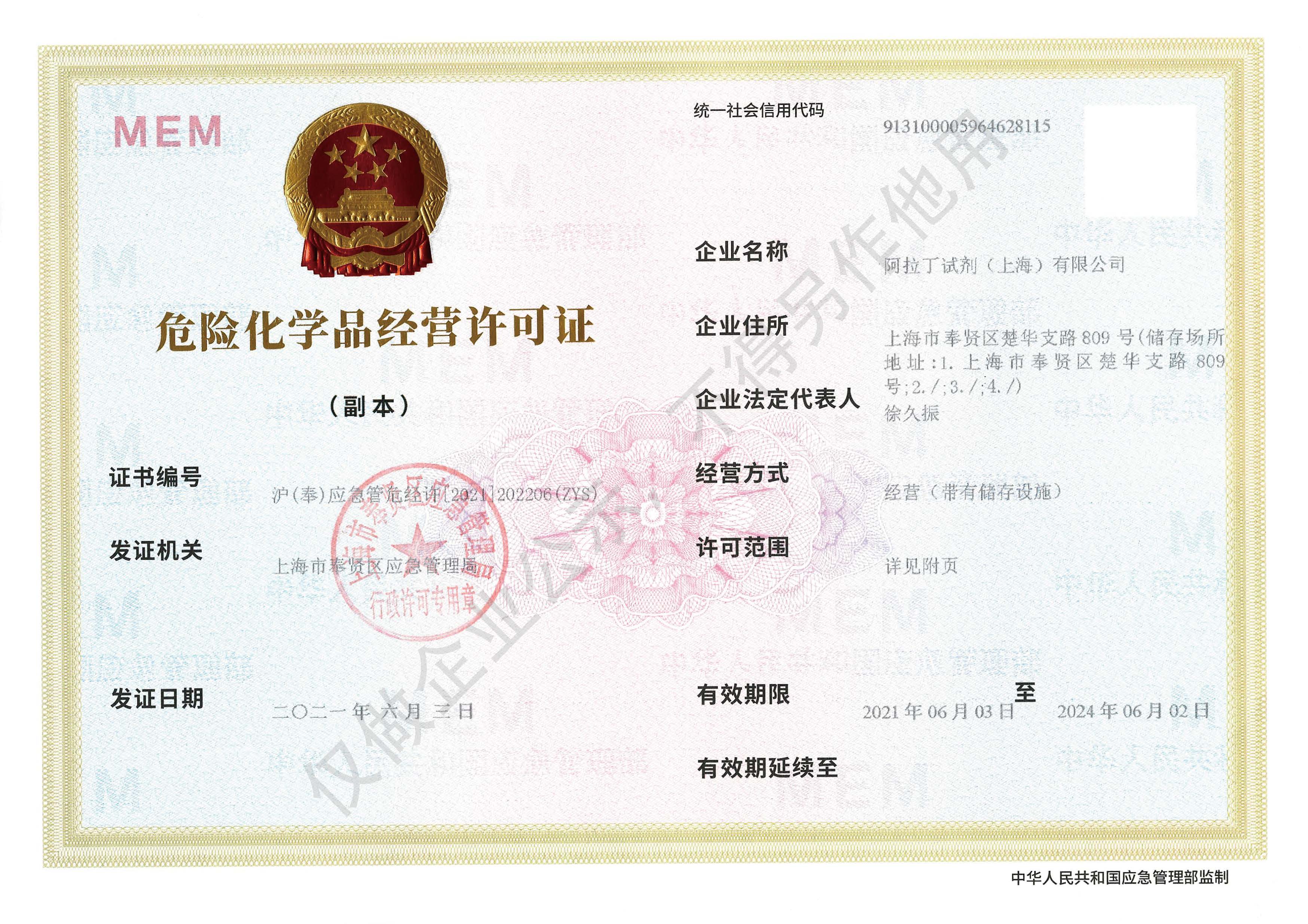 危险品化学品经营许可证(带存储)