 首页
首页 400-620-6333
Antibody structure and isotypes
Antibody structure?

Antibodies, also referred to as immunoglobulins (Ig), are significant Y-shaped glycoproteins produced by B-cells as a primary immune defense. These large proteins specially bind unique pathogen molecules that are known as antigens.
?
Antibodies are made up of one or more Y-shaped units consisting of four polypeptide chains?(Figure1). Each Y unit comprises two identical heavy chains (H) and two identical light chains (L), which differ in sequence and length. The top of the Y shape includes the variable region (V), also referred to as the fragment antigen-binding (F(ab)) region, which can firmly bind to a particular part of an antigen called an epitope.The antibody base is composed of constant domains (C) and generates the fragment crystallize?region (Fc), which is crucial for antibody function in an immune response.
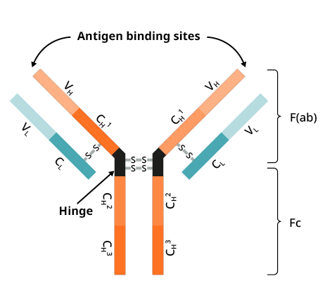
Figure 1. Antibody Structure. The Y-shaped antibody is held together at the center by a flexible hinge region. Antigen binding takes place at the variable domain (V), which comprises immunoglobulin heavy (H) and light (L) chains. The antibody's base contains constant domains (C). VH?refers to the heavy chain variable domain, VL refers to the light chain variable domain, CH refers to the heavy chain constant domain, and CL?refers to the light chain constant domain.
F(ab) and Fc regions
The proteolytic enzyme pepsin can cleave the Y-shape of an antibody into three fragments, specifically two F(ab) regions and an Fc region. The F(ab) regions comprise the variable domain, which binds selectively to antigens. The Fc fragment provides a binding site for endogenous Fc receptors on the surface of lymphocytes and secondary antibodies. In addition, dye and enzymes may be covalently linked to antibodies at the Fc portion for experimental visualization.
Antibody fragments offer distinct benefits in certain immunochemical techniques. Fragmenting IgG antibodies can be useful as F(ab) fragments (1) will not cause the antigen to precipitate and (2) will not bind to immune cells in in vivo studies due to the absence of an Fc region. F(ab) fragments are often used in functional studies because of their smaller size and lack of cross-linking (due to the loss of the Fc region). On the other hand, Fc fragments are commonly used as Fc receptor blockers in immunohistochemical staining.
Heavy chains
The heavy chain type determines the antibody's class or isotype. Mammalian Ig heavy chains can be classified into five types identified by : α, δ, ε, γ and?μ, which correspond to IgA, IgD, IgE, IgG, and IgM antibodies. Each heavy chain type differs in size and composition, α and γ chains contain about 450 amino acids, whereas μ and ε chains contain about 550 amino acids.
?
Each heavy chain has two regions: constant (CH) and variable (VH). The constant region is identical in isotype antibodies but differs in antibodies of different isotypes. Heavy chains γ, α, and δ consist of three tandem Ig domains?(CH1, CH2, CH3)?and a hinge region for added flexibility. Heavy chains μ and ε consist of four immunoglobulin domains. The variable region (VH) of the heavy chain differs depending on the producing B cell but remains the same for all antibodies produced by a single B cell or B cell clone. Each heavy chain's variable region is composed of a single Ig domain and is approximately 110 amino acids in length.
Light chains
Mammals have only two types of light chain, lambda (λ) and kappa (κ), which differ slightly in polypeptide sequence. A light chain comprises two consecutive domains: constant (CL) and variable (VL). The length of a light chain is approximately 211–217 amino acids. Antibodies contain two identical light chains. Additional types of light chains, for instance, the iota (ι) chain, exist in lower vertebrates such as Chondrichthyes and Teleostei.
Antibody isotypes
In mammals, antibodies are categorised into five isotypes: IgG, IgM, IgA, IgD, and IgE.?Each isotype boasts a unique structure represented in Figure 2. The isotypes differ depending on the quantity of Y units and heavy chain types. Distinctions can also be found in their biological properties, functional locations and capacity to handle diverse antigens, as shown in Table 1.
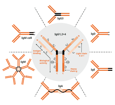
Figure 2.?Antibody structure and isotypes.
?
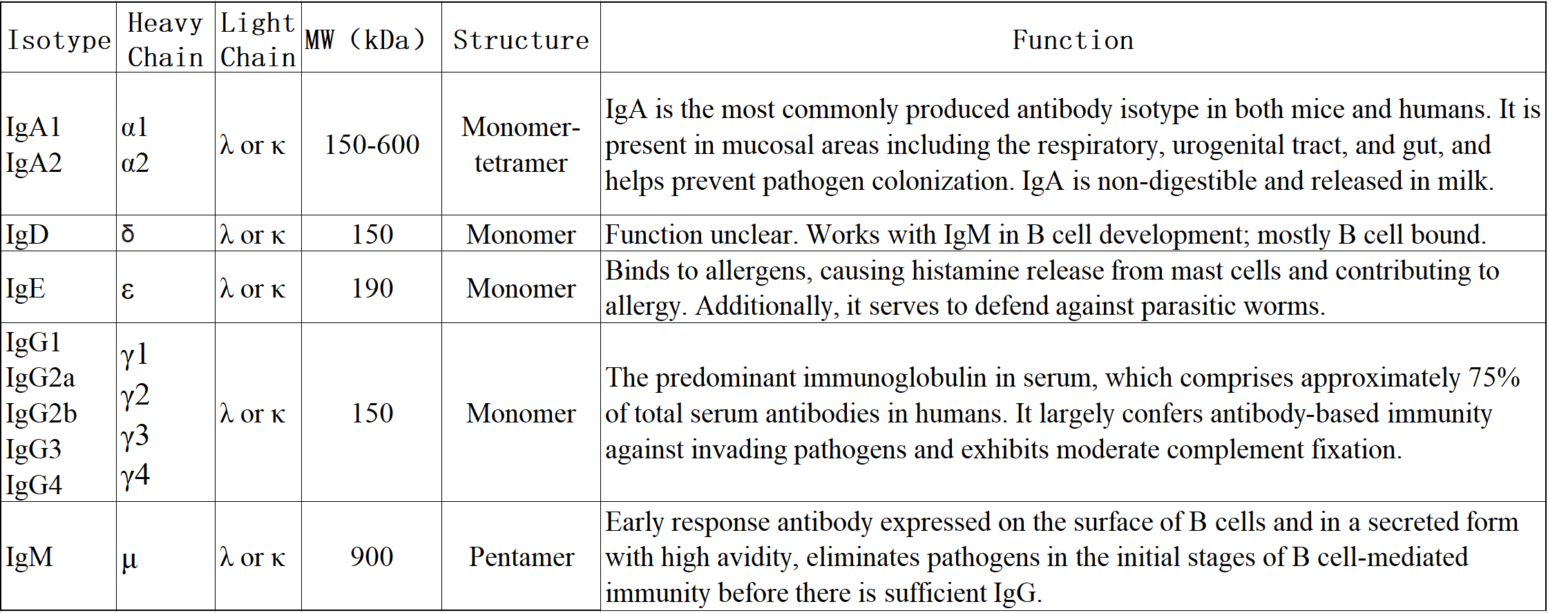
Table 1.?Structure and functions of different antibody isotypes.?
Antigen-antibody interactions: How and Where antibodies bind
The F(ab) antibody region includes the antigen-binding site, paratope. Paratope connects to a particular area of an antigen known as epitope, which is often only a few amino acids long (Fig. 3).
?
The paratope and epitope are held together by complementary shapes and intermolecular interactions such as Van der Waals, hydrogen bonds, electrostatic and hydrophobic interactions; the strength of these forces determines the affinity of the antibody.
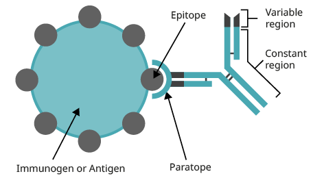
Figure 3.?A schematic representation of antigen-antibody interactions.
Reference
1.?Alberts B, Johnson A, Lewis J, et al. Molecular Biology of the Cell. 4th edition. New York: Garland Science; 2002. Chapter 24, The Adaptive Immune System. https://www.ncbi.nlm.nih.gov/books/NBK26830/
2.?Murphy K, Weaver C. Janeway's Immunobiology. 9th edition. New York: Garland Science; 2016. Chapter 5, Antibodies and Antigens.? https://www.ncbi.nlm.nih.gov/books/NBK27125/
Janeway CA, Travers P, Walport M, et al. Immunobiology: The Immune System in Health and Disease. 5th edition. New York: Garland Science; 2001. Chapter 5, Antibodies and Antigens. https://www.ncbi.nlm.nih.gov/books/NBK27101/


 危险品化学品经营许可证(带存储)