 首页
首页 400-620-6333
Silica-Coated Gold Nanoparticles: Surface Chemistry, Properties, Benefits, and Applications
Introduction
various fields ranging from biomedical engineering [1] to photovoltaic power generation [2].?Most applications take advantage of the excellent optical properties of gold nanoparticles, which can be fine-tuned by changing their shape and size.
?
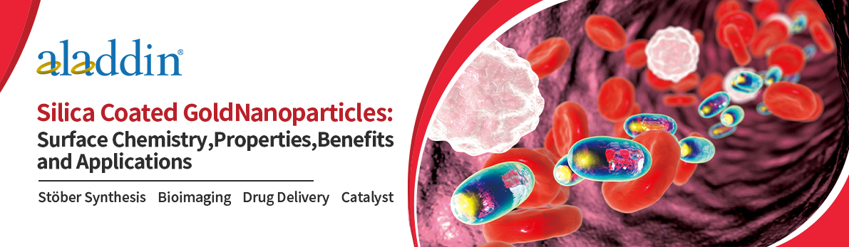
However, uncoated gold nanoparticles tend to aggregate in solution and melt under laser irradiation, both of which lead to significant changes in their optical properties. When their surfaces are properly passivated by chemical functionalization, they resist aggregation and shape changes under a wide range of biological, physical, and environmental conditions, thereby preserving their optical properties. One of the powerful functionalization methods that have?been shown to enhance the thermodynamic and chemical stability of gold nanoparticles is silica coating [1,3,4]. The excellent stability and functionality imparted by silica coatings make them the first choice for many of the applications described below. Examples of silica-coated gold nanospheres and nanorods are shown in Figure 1.
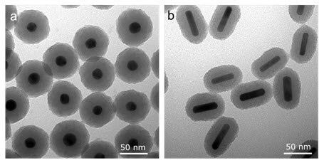
Figure 1: SEM image of silica-coated gold nanospheres and (b) SEM image of silica-coated gold nanorods
Surface Chemistry and Synthesis of Silica-Coated Gold Nanoparticles
The silica-coating of gold nanoparticles was accomplished using the classical St?ber method involving tetraethyl orthosilicate (TEOS), a highly branched and mesoporous siloxane polymer formed on the gold surface. The reaction can be controlled so that the thickness of the silica layer on the gold surface can be tailored based on reaction time and reagent concentration. The resulting surface siloxane polymer, known as silica, has hydroxyl (-OH) groups that can be used as a basis for further functionalization. In addition, heterobifunctional silane linkers react readily with silica, providing a means to attach various ligands, such as polyethylene glycol (PEG), to silica surfaces (Figure 2).
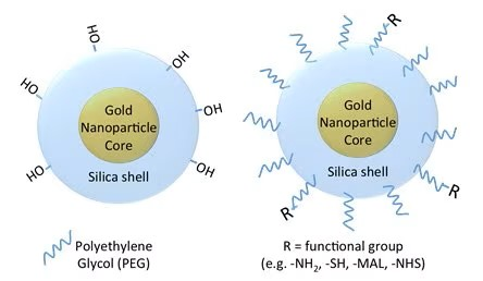
Figure 2: Surface chemistry of silica-coated gold nanoparticles. Shown are?a bare silica surface with hydroxyl functional groups (left) and a nanoparticle coated with polyethylene glycol (PEG) that can be functionalized with different end groups, including amine, thiol, maleimide and n-hydroxysuccinimide et al.
Properties and Advantages of Silica-Coated Gold Nanoparticles
Silica coatings of gold nanoparticles are advantageous in many applications involving gold nanoparticles. Especially in pulsed laser applications, the silica coating greatly enhances the thermodynamic stability of gold nanorods. Gold nanorods with standard coatings such as PEG, CTAB, or other small polymers can absorb enough energy from a pulsed laser to melt (Figure 3). This change in shape results in a corresponding change in the light it absorbs and scatters, from near-infrared wavelengths to the visible spectrum. For applications where consistent near-infrared light absorption is critical, gold nanorods with a silica coating are resistant to shape change And can maintain their?optical properties at higher light intensity levels (Figure 4).
Figure 3: Gold nanorods and their optical properties before (left) and after (right) exposure to pulsed laser light. Standard gold nanorods without a silica coating are very unstable and melt in response to laser absorption, reducing their absorption and scattering of light at near-infrared wavelengths.
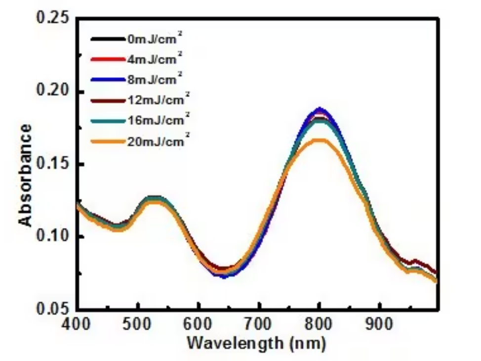
Figure 4: Absorbance spectra of silica-coated gold nanorods after exposure to 300 pulses of 808 nm light at different intensities (fluence). Silica-coated particles are thermodynamically stable (they resist shape changes) under influences of up to 20 mJ/ cm2.
In addition to thermodynamic stability (anti-melting) and colloidal stability, the silica coating of gold nanorods has other significant properties and advantages. For example, silica coatings increase the surface area for the conjugation of antibodies or other targeting moieties. In addition, silica is porous and can be loaded with drugs, dye molecules, or other imaging agents via physical adsorption or covalent attachment. The silica coating also limits how tightly packed the nanorods can be at high concentrations, marginalizing the effects of plasmonic coupling and allowing the preservation of concentration-independent optical properties. Finally, the silica coating on gold nanoparticles can increase the imaging contrast by more than 3 times [1].
PEGylated silica-coated gold nanoparticles in photoacoustic imaging (Figure 2-right) can provide all the benefits of silica-coating, as well as the performance associated with PEG-coated nanoparticles, including higher colloidal stability and lower immunogenicity.
Applications of Silica-Coated Gold Nanostructures
1.?Photoacoustic Imaging
When used as a contrast agent in photoacoustic imaging (photoacoustic imaging), the gold nanorods absorb light from a pulsed laser and generate a lot of heat. While this heat is required for the photoacoustic effect, too much heat is inappropriate, causing the nanorods to melt (Figure 2). This shape change reduces?the absorption cross-section, which leads to a loss of contrast in photoacoustic imaging. The silica coating helps reduce the interfacial thermal resistance between the gold and the surrounding solvent (Fig. 5), allowing the particle to release more heat to its environment, which has two positive effects: (1) the gold particle can Resistance to melting at high throughput (Fig. 3), (2) thermodynamic stability of the photoacoustic signal generated by these particles is at least 3 times greater than that of gold nanoparticles with standard coatings such as PEG, CTAB or other small polymers [1].
Thermodynamically stable silicon-coated gold nanorods and nanospheres have become popular for photoacoustic contrast agents and therapeutic agents due to their superior thermodynamic stability, optical properties, biocompatibility, and bioconjugation potential [1].

Figure 5: Schematic representation of the heat transport process from a nanoparticle to the environment, and the resulting temporal distribution of the temperature (T) near the nanoparticle surface and the amplitude of the photoacoustic signal (P) away from the surface. (a) Bare nanoparticles with high interfacial resistance lead to broadened temperature distribution and smaller amplitude of photoacoustic pressure signal; (b) introduction of silicon shell minimizes interfacial resistance between gold (Au) and SiO2?and?water. The resulting sharper temperature profile, and since the temperature profile is over a greater distance, the photoacoustic signal increases; (c) the thicker shell causes the temperature peak to broaden, and the photoacoustic signal drops again, although it may still be higher than the bare nanoparticles.
2.?Cell Tracking
Since photoacoustic imaging (PAI) is non-invasive, quantitative, and has a short scan time, it is an ideal tool for immune cell tracking [8]?and stems?cell engraftment tracking [10]?in combination with ultrasound imaging?(Fig.6). Silica-coated gold nanorods can be used as contrast agents in photoacoustic imaging to quantify implanted cells in real-time?and confirm that sufficient numbers of cells have reached the treatment site. The silica coating also helps to promote the cellular uptake of gold nanorods [8].
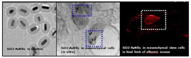
Figure 6: The first image shows a TEM image of SiO2 -AuNRs (silica-coated gold nanorods) with a peak absorption at 676 nm and a silica coating thickness of 20 nm, and the second image confirms that?There are SiO2-AuNRs?in murine mesenchymal stem cells; the third figure shows contrast-enhanced photoacoustic images of SiO2-AuNRs-labeled mesenchymal stem cells injected intramuscularly into the hindlimb of a mouse with a developing thymus.
Silica-coated gold nanorods are also widely used in photothermal therapy due to their small size, tunable resonances in the red and near-infrared spectra, and very high absorption cross-sections. The silica coating increases the photothermal stability of gold nanorods, thereby helping to maintain their superior optical properties under high continuous and pulsed laser fluences [1,3,6]. Silica-coated gold nanorods also showed significantly increased cellular uptake than PEG-coated gold nanorods, which in turn translated into better photothermal ablation effects [10]. Furthermore, silica-coated nanorods can be used in conjunction with photoacoustic imaging to simultaneously create a map of heat generation during photothermal therapy, which can guide its dosage and therapeutic effect [7].
3.?Targeted Drug Delivery
The silica-coated gold nanoparticles are biocompatible and can be chemically modified to specifically target cancerous tissue. Mesoporous silica-coated gold nanoparticles enable chemical modification due to their large surface area, tunable size, high accessible pore volume, drug loading capacity, and well-defined surface properties (Fig. 4). Compared with photothermal therapy and chemotherapy alone, combined chemo photothermal?therapy completed by drug-loaded silica-coated gold nanorods has been shown to have enhanced anticancer effects [11].
4.?Multiple Images
Targeted silica-coated gold nanorods can be used as contrast agents for multiplex imaging to distinguish cellular inclusions in vitro by targeting nanoparticles to cells expressing different cellular receptors[12] (Fig.7). Silica-coated gold nanorods with different peak wavelengths can be used to label each unique cell type to locate specific cell types and generate images corresponding to molecular expression.
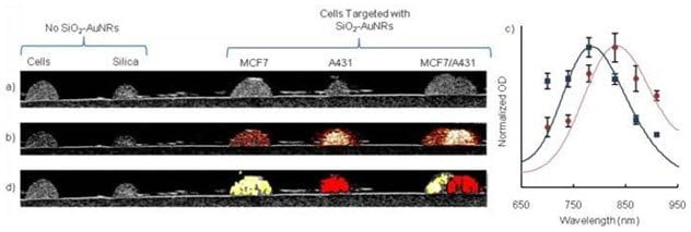
Figure 7: Signal processing and statistical analysis of cell phantom photoacoustic (PA) images confirm the unique identification of cellular inclusions: a) inclusions are visible in ultrasound images; b) at 830?The PA image acquired at nm shows silicon-coated gold nanorods ( SiO2-AuNR ) in inclusions; c) Comparison of PA signal intensity (dots) and UV-VIS spectrum (solid line) shows that SiO2-AuNR light absorption spectrum determines the PA signal intensity. The inclusions were segmented into three regions and the PA signal intensities were averaged; d) Molecular map of the cell and US overlay; 830 nm silica-coated gold nanorods are shown in red, and 780 nm SiO2-AuNR?are shown in yellow.
5.?Dual Mode/Multimodal Imaging
Although optical imaging techniques have high sensitivity and can visualize diseased tissue with excellent resolution, they are limited by the low penetration of light in tissue. Using a combination of complementary imaging modalities can help optimize sensitivity and specificity. For example, contrast agents that simultaneously enhance CT contrast and near-infrared optical imaging can provide quantifiable information on the accumulation of contrast agents at different levels. Mesoporous silicon-coated gold nanorods loaded with organic NIR dyes such as indocyanine green (ICG) can be used as imaging probes for dual-mode X-ray CT and NIR fluorescence imaging [13].
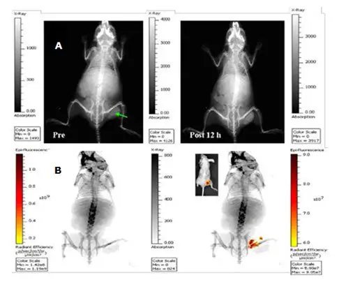
X-ray?images of mice before and 12 h after intratumoral injection of indocyanine green-loaded silica-coated gold nanorods (200 μL, 1.5 mg/mL) (exposure time 30 s); (b) In vivo planar X-ray?image with 60 s exposure time (left) superimposed with the corresponding near-infrared fluorescence image (10 s exposure time) (right) after intratumoral injection of dual-mode imaging contrast agent for 12 h. Inset: corresponding overlay of bright-field and near-infrared fluorescence images. Green arrows indicate tumors. A thin layer of silica sandwiched between the gold nanorods and the indocyanine green chromophore protects the dye from fluorescence quenching.
6.?Surface Enhanced Raman Spectroscopy (SERS)
Dye-embedded silica-coated gold nanoparticles are very effective for surface Raman enhancement and can be used as multiplex detection and spectroscopic labeling particles [14]. Metallic cores for optical signal enhancement and signature molecules for spectral labeling are composed of silica shells.

Figure 9: Schematic diagram of the structure of core-shell nanoparticles and the process of preparing silica-coated SERS active gold colloids. (a) Under the excitation of 632 ~ 647 nm wavelength, the size range of colloidal gold particles optimized for surface Raman enhancement is 55 ~ 65nm; (b) gold particles with adsorbed Raman reporter factors; (c) containing Gold particles with reporter and mercaptopropyl trimethoxysilane, a common coupling agent; (d) silica-coated gold particles with a Raman spectroscopic reporter embedded at the core-shell boundary.
Silica-coated gold nanorods have also been used as dual-modal imaging probes targeting cancer cells. Fluorescence and surface-enhanced Raman scattering signals can be independently displayed by different nanoparticle excitation wavelengths [17].
7.?Two-Photon Imaging
Mesoporous silicon-coated gold nanorods containing photosensitizers such as Pd-mesoporous tetrakis(4-carboxyphenyl)porphyrins (PdTPPs) can be used for two-photon-activated photodynamic therapy (Fig. 10) [16]. Doping photosensitizers into the nanochannels of the mesoporous silica shell can excite photosensitizers through the intraparticle plasmon resonance energy transfer of encapsulated two-photon excited gold nanorods, and can generate cytotoxic singlet oxygen to kill dead cancer cells. The silica matrix can significantly enhance the two-photon emission stability of the gold nanorod core by providing mechanical support against thermal deformation.
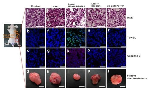
Figure 10: In vivo studies demonstrating two-photon-activated photodynamic therapy following intratumoral injection of mesoporous silicon-coated gold nanorods PdTPPs. Stained with hematoxylin and eosin (first row), TUNEL (second row, green), Caspase -3 (third row, red),?and DAPI (blue), against irradiation for 24?After h, tumor sections were harvested for histological analysis. The inhibitory effect of mesoporous silicon-coated gold nanowires-PdTPPs combined with laser irradiation on tumor growth was observed by apoptosis indicators TUNEL and Caspase-3 staining (1.2 times, Figure 6i).
8.?Biomolecular Probes
Due to the high emissivity of silica in the mid-infrared range, silica-coated gold nanoparticles can be used as
Labeling of DNA hybridization assays to detect lower target DNA concentrations than citrate-coated or standard gold nanoparticles. Fluorescence-based DNA hybridization and protein-binding assays can be improved using silicon-coated gold nanoparticles as robust labels that do not degrade, quench, or photobleach[17]. This nanoparticle platform can also be used to detect small molecules such as proteins, bacteria, pesticides,?and mercury at very low concentrations.
9.?Catalyst
For gold nanoparticles to be effective catalysts, they must be highly stable in the surrounding medium, and thermal environment, and have recycling potential while maintaining catalytic activity. Typically, gold nanorods are coated with a surfactant called cetyltrimethylammonium bromide (CTAB). In organic solutions, desorption of CTAB into the medium would lead to the aggregation of gold nanorods and loss of their catalytic properties. In addition, CTAB-coated gold nanorods may be thermodynamically unstable, which can induce transformation into spherical particles. Mesoporous silica-coated gold nanorods are very effective as catalysts because the silica shell provides high stability under adverse conditions including heat, solvent exchange, and centrifugation [18]. Due to the large pore volume of the mesoporous silica shell, the reactant molecules can diffuse through the pores to the gold surface for catalysis.
10.?Photonics
Plasmonic gold nanoparticles are used in many photonics applications; these include single-molecule detection, photonic crystal construction, and optical device design (such as waveguides). However, the formation of the photonic bandgap is prevented due to the physical contact between the metals of adjacent bare gold nanoparticles. Coating gold nanoparticles with a layer of optically transparent, chemically inert, and photochemically stable materials such as silicon dioxide, a complete photonic bandgap can be generated when plasmonic nanoparticles are organized in a periodic structure [19]. In addition, the silica coating also contributes to plasmonic fluorescence enhancement because it suppresses the quenching mechanism that occurs when fluorophores bind directly to metal surfaces. Thus, silica-coated gold nanoparticles offer several advantages over standard gold nanoparticles.
?
In summary, silica-coated gold nanoparticles can provide several beneficial properties, including:
● Higher imaging signal intensity Superior thermal and colloidal stability in photoacoustic imaging and other modalities involving the use of pulsed lasers;
● Stable optical properties for a wider range of applications, including multi-modal imaging therapeutic capabilities for use as hybrid drug carriers and imaging agents;
● Flexible silane chemistry for covalent functionalization and coupling.
These unique advantages make them ideal for many applications.
References
1.Chen Y, Frey W, Kim S, Kruizinga P, Homan K, Emelianov S. 2011. Silica-Coated Gold Nanorods as Photoacoustic Signal Nanoamplifiers. Nano Lett.. 11(2):348-354. https://doi.org/10.1021/nl1042006
2. Yang J, You J, Chen C, Hsu W, Tan H, Zhang XW, Hong Z, Yang Y. 2011. Plasmonic Polymer Tandem Solar Cell. ACS Nano. 5(8):6210-6217. https://doi.org/10.1021/nn202144b
3.Chen Y, Frey W, Kim S, Homan K, Kruizinga P, Sokolov K, Emelianov S. 2010. Enhanced thermal stability of silica-coated ?gold nanorods for photoacoustic imaging and image-guided therapy. Opt. Express. 18(9):8867. https://doi.org/10.1364/oe.18.008867
4.Luke GP, Bashyam A, Homan KA, Makhija S, Chen Y, Emelianov SY. 2013. Silica-coated gold nanoplates as stable photoacoustic contrast agents for sentinel lymph node imaging. Nanotechnology.?24(45):455101. https://doi.org/10.1088/0957-4484/24/45/455101
5.Zijlstra P, Chon JWM, Gu M. 2009. Five-dimensional optical recording mediated by surface plasmons in gold nanorods. Nature. 459(7245):410-413. https://doi.org/10.1038/nature08053
6. Kim S, Chen Y, Luke GP, Emelianov SY. 2014. In-vivo ultrasound and photoacoustic image-guided photothermal cancer therapy using silica-coated gold nanorods. IEEE Trans. Ultrason., Ferroelect., Freq. Contr.. 61(5):891-897. https://doi.org/10.1109/tuffc.2014.2980
7. Chen Y, Frey W, Walker C, Aglyamov S, Emelianov S. 2013. Sensitivity-enhanced nano thermal sensors for photoacoustic temperature mapping. J. Biophoton.. 6(6-7):534-542. https://doi.org/10.1002/jbio.201200219
8.Joshi PP, Yoon SJ, Chen Y, Emelianov S, Sokolov KV. 2013. Development and optimization of near-IR contrast agents for immune cell tracking. Biomed. Opt.?Express. 4(11):2609. https://doi.org/10.1364/boe.4.002609
9. Jokerst JV, Thangaraj M, Kempen PJ, Sinclair R, Gambhir SS. 2012. Photoacoustic Imaging of Mesenchymal Stem Cells in Living Mice via Silica-Coated Gold Nanorods. ACS Nano. 6(7):5920-5930. https://doi.org/10.1021/nn302042y
10.Zhu X, Fang C, Jia H, Huang Y, Cheng CHK, Ko C, Chen Z, Wang J, Wang YJ. Cellular uptake behavior, photothermal therapy performance, and cytotoxicity of gold nanorods with various coatings. Nanoscale. 6(19):11462-11472. https://doi.org/10.1039/c4nr03865g
11. Monem AS, Elbialy N, Mohamed N. 2014. Mesoporous silica-coated gold nanorods loaded doxorubicin for combined chemo?photothermal therapy. International Journal of Pharmaceutics. 470(1-2):1-7. https://doi.org/10.1016/j.ijpharm.2014.04.067
12.Bayer CL, Chen Y, Kim S, Mallidi S, Sokolov K, Emelianov S. 2011. Multiplex photoacoustic molecular imaging using targeted silica-coated gold nanorods. Biomed. Opt. Express. 2(7):1828. https://doi.org/10.1364/boe.2.001828
13. Luo T, Huang P, Gao G, Shen G, Fu S, Cui D, Zhou C, Ren Q. 2011. Mesoporous silica-coated gold nanorods with embedded indocyanine green for dual mode X-ray CT and NIR fluorescence imaging. Opt. Express. 19(18):17030. https://doi.org/10.1364/oe.19.017030
14.Doering WE, Nie S. 2003. Spectroscopic Tags Using Dye-Embedded Nanoparticles and Surface-Enhanced Raman Scattering. Anal. Chem.. 75(22):6171-6176. https://doi.org/10.1021/ac034672u
15.Wang Z, Zong S, Yang J, Li J, Cui Y. 2011. Dual-mode probe based on mesoporous silica coated gold nanorods for targeting cancer cells. Biosensors and Bioelectronics. 26(6):2883-2889. https://doi.org/10.1016/j.bios.2010.11.032
16.Chen N, Tang K, Chung M, Cheng S, Huang C, Chu C, Chou P, Souris JS, Chen C, Mou C, et al. 2014. Enhanced Plasmonic Resonance Energy Transfer in Mesoporous Silica-Encased Gold Nanorod for Two-Photon-Activated Photodynamic Therapy. Theranostics.?4(8):798-807. https://doi.org/10.7150/thno.8934
17.Xia F, Zuo X, Yang R, Xiao Y, Kang D, Vallee-Belisle A, Gong X, Yuen JD, Hsu BBY, Heeger AJ, et al. 2010. Colorimetric detection of DNA, small molecules, proteins, and ions using unmodified gold nanoparticles and conjugated polyelectrolytes. Proceedings of the National Academy of Sciences. 107(24):10837-10841. https://doi.org/10.1073/pnas.1005632107
18.Son M, Lee J, Jang D. 2014. Light-treated silica-coated gold nanorods have highly enhanced catalytic performances and reusability. Journal of Molecular Catalysis A: Chemical. 38538-45. https://doi.org/10.1016/j.molcata.2014.01.010
19.Rodríguez-Fernández J, Pastoriza-Santos I, Pérez-Juste J, García de Abajo FJ, Liz-Marzán LM. 2007. The Effect of Silica Coating on the Optical Response of Sub-micrometer Gold Spheres. J. Phys. Chem. C.?111(36):13361-13366. https://doi.org/10.1021/jp073853n


 危险品化学品经营许可证(带存储)