 首页
首页 400-620-6333
Protein sample preparation process
1、Protein extraction
The first step of protein analysis is protein extraction, which includes chemical extraction, physical extraction and biological extraction. Protein extraction, whether through mechanical treatment or based on detergents, will inevitably destroy cell homeostasis, leading to protein degradation or instability. Therefore, the integrity of protein in the extraction process directly determines the quality of data obtained from the analysis of downstream protein samples.
Sample type
The research sources of endogenous proteins and expression system proteins mainly include mammalian cells, mammalian tissues and primary cells. When extracting proteins from mammalian tissues, some mild enzymes or mechanical crushing methods are needed to help isolate cells from more complex tissue matrix. For the cultured mammalian cells and primary cells that separate the cell contents from the environment only through the plasma membrane, the detergent in the reagent can break the protein-lipid membrane bilayer structure, making it easier to extract the total protein. Other organisms commonly studied or used for recombinant protein expression systems include bacteria, yeast and plants. These cell types contain cell walls and require additional enzymatic hydrolysis or mechanical fragmentation to effectively release proteins. However, solutions based on detergents have been developed, which can effectively extract and dissolve the proteins in these cells without mechanical damage. This method is particularly important for processing more samples or using automatic extraction and purification schemes.
Subcellular fractionation
For most studies, it is only necessary to directly prepare whole-cell lysates to obtain soluble protein samples for downstream direct detection or further purification and separation. However, if cells are divided into different compartments or organelles before protein extraction, the yield or enrichment rate of specific proteins can be significantly increased. Mechanical lysis usually destroys all cell compartments, making it more difficult to separate specific cell components. However, through careful optimization of reagents, a method based on stepwise differential detergent has been developed to separate nuclear protein, cytoplasmic protein and membrane protein components. This method can dissolve hydrophobic membrane proteins and separate them from hydrophilic proteins. It can also separate complete nuclei, mitochondria and other organelles for direct research or step by step extraction of proteins.
2、Protein degradation
During cell lysis, cell membrane and organelles will be destroyed, resulting in uncontrolled proteolytic activity, thus reducing protein production and affecting protein function. In order to prevent the degradation of the extracted protein and maintain its activity, it is usually necessary to add protease and phosphatase inhibitor to the cell lysis reagent.
Protease inhibitors act by binding to the active site of protease. Due to the difference of protein hydrolysis mechanism, a single compound cannot effectively inhibit all proteases. Therefore, it is necessary to use a mixture of several different inhibitors to ensure that the protein extract is not degraded before downstream analysis. Typical inhibitor mixtures include small molecule inhibitors such as serine, cysteine and aspartate protease, as well as aminopeptidase and metalloproteinase. Although some inhibitors are irreversible, many of them are reversible. They need to exist in crude samples for a long time until they are subsequently purified to eliminate the risk of proteolytic activity.
Similarly, phosphatases are different, so it is recommended to use effective phosphatase mixtures (inhibitors containing serine, threonine, tyrosine, acid and alkaline phosphatase) to maintain weak phosphorylation post-translational modification.
Prevent target protein degradation by using the correct preservation method:
Operate quickly and keep the sample low temperature (liquid nitrogen can be used to freeze the sample);
Inhibit or inactivate endogenous protease and phosphatase;
Adding protective or stabilizing compounds, such as reducing agents and enzyme inhibitors;
Stabilize or inactivate proteins by precipitation.
3、Protein sample impurity removal
After protein extraction, protein samples usually contain impurities that affect protein stability or are incompatible with downstream applications. Dialysis, desalting and concentration are three common methods to remove common small molecular pollutants (such as salt and detergent) in protein samples. According to the downstream application requirements, the factors to be considered in selecting the method may include the initial amount of sample, protein function and processing time. There are various specifications and packaging forms for dialysis, desalination and concentration.
Dialysis
Dialysis is a classical separation technology, which removes small molecules and unwanted compounds in protein solution through selective diffusion of semi-permeable membrane. Place the sample and buffer on the opposite side of the membrane. Proteins larger than membrane pores remain on the sample side of the membrane, and smaller molecules (pollutants) diffuse freely through the membrane until the equilibrium concentration is reached. This technology can reduce the concentration of small molecular pollutants in the sample to an acceptable level.
Desalination
Common desalting methods include dialysis, electrodialysis and gel filtration. Dialysis and electrodialysis take a long time, sample dilution is large, and it is not easy to scale up for large-scale production, so they are rarely used in industrial production. In the process of gel filtration chromatography desalination, the size of salt molecules and protein molecules is very different. The salt molecules of small molecules in the protein solution enter the stationary phase with small pore size along with the chromatographic mobile phase, making their migration rate in chromatography small. However, because of the large molecular size, proteins cannot enter the stationary phase along with the mobile phase, so the migration rate in the chromatographic column is large. First, they flow out of chromatography to achieve desalination.
Concentrate
Protein concentration is similar to dialysis method, which uses semi-permeable membrane to separate protein from small molecular weight compounds. Unlike the passive diffusion principle of dialysis, concentration is achieved by centrifuging the solution through the membrane. In the process of centrifugation, the buffer solution and small molecular weight solute are passively passed through the membrane and collected on the other side (filtrate). Macromolecules (proteins) remain on the sample side of the membrane, and they are concentrated into small volume liquid (intercepted liquid) as the reagent passes through the membrane passively to the other side.
4、Protein quantification
Ultraviolet spectrophotometry
There are tyrosine and tryptophan containing conjugated double bonds in the protein molecule, which makes the protein have the maximum absorption value to the 280nm light wave. Within a certain range, the absorption value of the protein solution is proportional to its concentration, which can be used for quantitative determination. The method is simple and fast, and the determined samples can be recovered, and the low concentration salt does not interfere with the determination results, so it is widely used in the biochemical preparation of protein and enzyme. However, this method has the following disadvantages:
1. When the contents of tyrosine and tryptophan residues in the protein to be measured differ greatly, certain errors will occur, so this method is applicable to the determination of samples with similar amino acid composition to the standard protein. ?
2. If the sample contains other substances absorbed at 280nm, such as nucleic acid and other compounds, there will be greater interference. However, the absorption peak of nucleic acid is 260 nm, so the light absorption values at 280 nm and 260 nm are measured respectively. The interference of nucleic acid on the determination of protein concentration can be properly eliminated through calculation. However, because the ultraviolet absorption of different proteins and nucleic acids is different, there are still some errors in the determination results after correction.
Fluorescence protein quantification
Fluorescence based protein quantification is an alternative to colorimetry. The fluorescence detection method has excellent sensitivity and requires less protein samples, which can save more samples for other experiments. In addition, reading time is not a key factor, so this detection method can be easily applied to automated high-throughput analysis. Fluorescence signal can be detected by fluorescence meter or enzyme marker.
Bradford method
In 1976, Bradford established the principle of combining protein with Coomassie Brilliant Blue G-250, which is a rapid and accurate method to quantify protein. The combination of dye and protein causes the change of maximum absorption light of dye from 465 nm to 595 nm. The protein-dye complex has a high extinction coefficient, which greatly improves the sensitivity of protein determination (the minimum detection amount is 1 μ g)。 The combination of dye and protein is a very rapid process, only takes about 2 minutes, and the color of the complex is stable within 1 hour. Some cations such as K+, Na+, Mg2+, (NH4) 2SO4, ethanol and other substances do not interfere with the determination, while a large number of detergents such as TritonX-100 and SDS seriously interfere with the determination, and a small amount of detergents can be eliminated by using appropriate control. Because of its simplicity and rapidity, less interfering substances and high sensitivity, the staining method has been widely used for the determination of protein content.
Determination of total nitrogen content - micro-Kjeldahl method: when the measured nitrogenous organic matter is co-digested with concentrated sulfuric acid, nitrogen, carbon dioxide and water are decomposed, and the nitrogen and sulfuric acid are combined to form ammonium sulfate. Because the decomposition reaction proceeds very slowly, copper sulfate and potassium sulfate or sodium sulfate can be added, in which copper sulfate is the catalyst, potassium sulfate or sodium sulfate can improve the boiling point of the digestion solution, and oxidant (hydrogen peroxide) can also accelerate the reaction.
After digestion, add strong alkaline alkaline digestion solution into the Kjeldahl nitrogen analyzer to release ammonia from the ammonia sulfate; Steam distillation is used to steam ammonia into excess standard inorganic acid solution. After all the ammonia is evaporated, titrate the collected amount of ammonia with standard hydrochloric acid solution, accurately measure the amount of ammonia, and then convert the protein content.
Biuret method
Compounds with two or more peptide bonds have biuret reaction. Protein can complexe with Cu2+in alkaline solution to form purplish red, and the color depth is proportional to the protein concentration. Therefore, colorimetric method can be used for determination, and the protein concentration can be determined by calculation according to the standard curve.
Folin-phenol reagent method
The classical method for determining protein content is developed on the basis of biuret method. It is simple, rapid and highly sensitive, 100 times more sensitive than the biuret method. The reagent used in Folin-phenol method consists of two parts, and reagent A is equivalent to biuret reagent. The peptide bond in protein reacts with alkaline copper sulfate in reagent A to form a copper-protein complex. This complex can undergo oxidation-reduction reaction with phosphomolybdic acid and phosphotungstic acid in reagent B. Because phosphomolybdic acid and phosphotungstic acid are easily reduced by phenolic compounds, they react blue. However, tyrosine and tryptophan in protein can undergo this color reaction. The depth of color is proportional to the concentration of protein. Therefore, the content of protein can be determined by colorimetry.
This method is easily disturbed by phenolic compounds and citric acid in protein samples. In addition, phosphomolybdic acid-phosphotungstic acid in reagent B is stable only at acidic pH value, so when adding reagent B to alkaline copper-protein solution, it must be mixed evenly immediately. To ensure that the reduction reaction can occur normally. This method is also applicable to the quantitative determination of tyrosine and tryptophan.
5、Protein detection
According to the experimental needs, many methods can be used to detect and analyze target proteins. The following are common technologies used to detect and analyze proteins in complex mixtures (such as lysates and serum), as well as the characteristics and common requirements of various technologies.
ELISA | western blot | Mass spectrometric analysis | |
superiority | High-throughput analysis of 96-well or 384-well plate can be performed to quantify target protein | Molecular weight determination and protein identification can separate and distinguish target proteins | Qualitative and quantitative analysis of multiple targets in the same sample to detect post-translational modification or different subtypes of protein |
sensitivity | <5–10 pg/mL | From low Feike to high Ake | Molar range (1018) |
Cracking fluid compatibility | For inactive detection ELISA: lysate based on ionic detergent For activity detection ELISA: cracking liquid based on non-ionic detergent (such as NP-40, Triton X-100) | For SDS-PAGE (denaturation): RIPA or cracking liquid containing other ionic detergent For non denaturing polyacrylamide gel electrophoresis (Native PAGE): pyrolysis liquid based on non-ionic detergent (such as NP-40, Triton X-100) | The detergent and high-salt substances in the sample must be removed before mass spectrometry analysis |
Conventional protein requirement | 0.1–1 μg/mL | 1–50 μg | <1 μg |
Required equipment | Microplate Reader | X-ray film or CCD imaging system | mass analyzer |


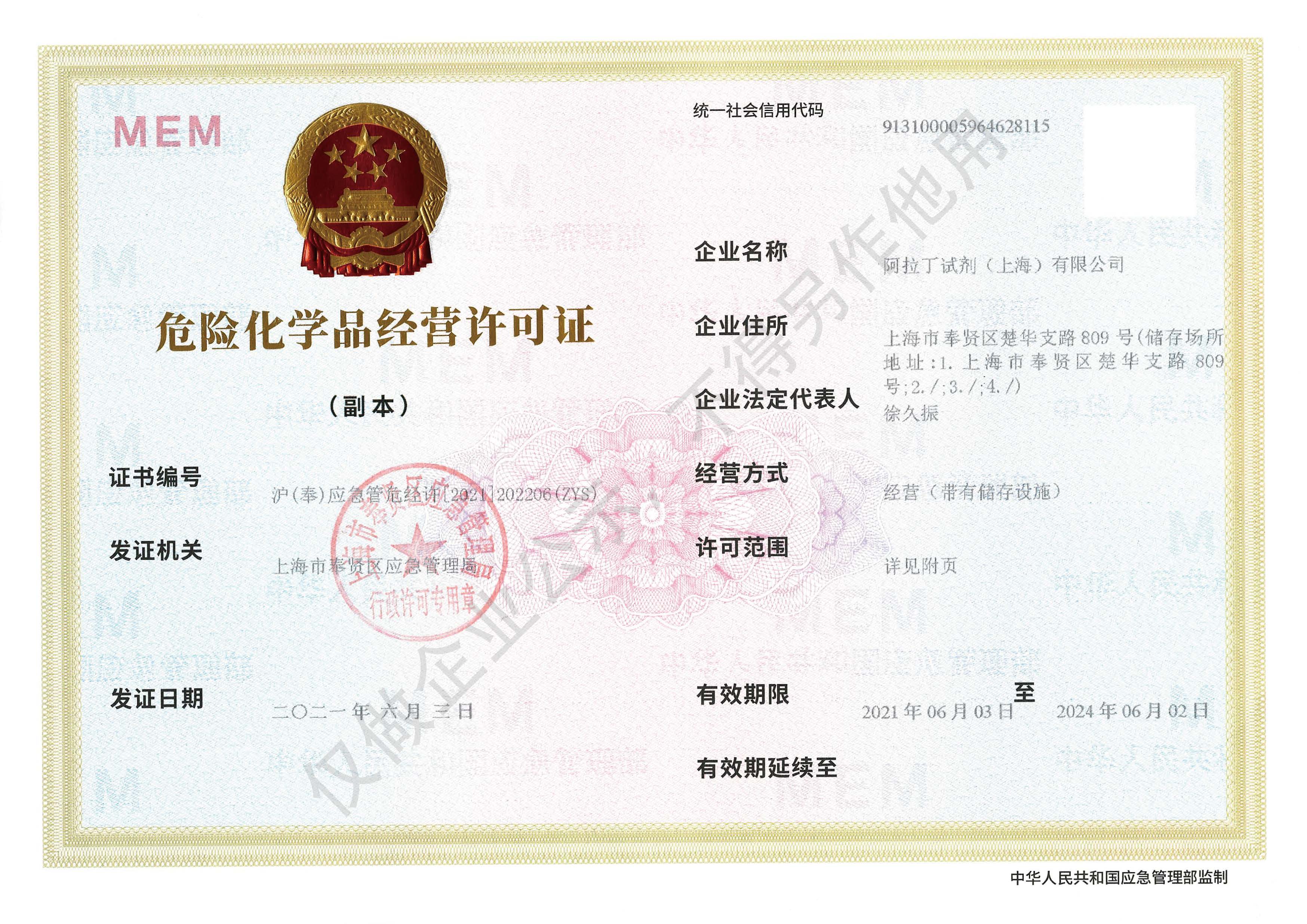 危险品化学品经营许可证(带存储)