 首页
首页 400-620-6333
Nanoparticle-Based Gene Delivery
In gene delivery, vectors are essential in the passage of DNA (hydrophilic, negatively charged) through cell membranes (hydrophobic, negatively charged). In addition, therapeutic efficiency also depends on the efficient delivery of DNA to the target site. Due to the many delivery barriers in the system, such as intracellular uptake, endosomal escape, DNA release, and nuclear uptake, etc., as well as extracellular barriers, such as avoiding particle clearance mechanisms, targeting specific tissues and/or cells of interest, protecting DNA is protected from degradation, etc., so developing an efficient and biocompatible gene delivery system is very important. So far, gene delivery systems can be divided into two categories, namely viral transduction systems and non-viral transduction systems. Although viruses can be used for gene delivery, the shortcomings of large DNA carried by them, low loading capacity, large-scale manufacturing, cost of quality control, immunogenicity, and potential carcinogenicity limit the application of viral vectors in gene delivery.
As a result, researchers have turned their attention to developing nonviral vectors as alternative vehicles for delivering genes. Non-viral delivery systems have several distinct advantages, including ease of preparation, synthetic manipulations suitable for polymer properties, cell or tissue targeting, low immunogenicity, and carcinogenicity, no viral recombination, and no limitation of the size of the carried DNA. , low manufacturing cost. In addition, non-viral vectors can easily deliver genetic material to target cells due to the advantages of their size, charge, and structurally modified vectors.
Nanomaterials are ideal materials for gene delivery because their physical properties make them suitable for specific functions. Inorganic nanomaterials are highly sought after due to their ease of functionalization, unique electrical and optical properties, biocompatibility, and low cytotoxicity. Magnetic nanoparticles, gold nanoparticles, quantum dots, and carbon nanotubes are commonly used inorganic materials for gene delivery.
Magnetic Nanoparticles
Magnetoinfection is a procedure based on the association of magnetic nanoparticles (MNPs) with gene carriers, which can enhance gene transfer in the presence of magnetic fields. It was originally developed by Christian Plank and colleagues who transferred genes in cell culture and in vivo by using MNPs-DNA complexes or MNPs-viral-vector complexes. The process is simple: add the MNPs - DNA complexes to a culture of adherent cells and place a magnet near the bottom of the flask or plate, attracting the magnetic complexes to the bottom where they come into close contact with the cells and are physically internalized, without any specific magnetic influence on the endocytic uptake mechanism. In vivo, adding a magnetic field at the target site increases the transfection yield and targets the therapeutic gene to a specific organ /location in the body. Generally, we inject particles with the therapeutic gene intravenously and apply a high gradient of external magnets at the target site to capture the particles. Once captured, the particles are retained at the target site and then absorbed by the tissue.
Gold Nanoparticles
Gold nanoparticles (AuNPs) are stable, uniform, and biocompatible metallic nanoparticles with unique electronic structures, size-dependent intensity displays, and highly tunable electronic, magnetic, and optoelectronic properties, making them a promising candidate for genetic Ideal for passing on. Furthermore, the soft surface chemistry of AuNPs enables them to be tailored with various biomolecules and ligands. For example, Klibanov et al. covalently attached ~2 kDa polyethyleneimine (PEI) chains to AuNPs to deliver plasmid DNA vectors into mammalian cells. They found that the most potent conjugate was 12-fold more efficient at delivering plasmid DNA than the unmodified PEI counterpart (Figure 1). Another important feature of AuNPs is their metallic core building blocks. This structure provides solid support for the therapeutic materials, making them stable even after infinite dilution.

Figure 1: Extent of β-gal gene expression mediated by PEI2 (panels ac) and PEI2-genii (panels d–f) in COS-7 cell cultures in the presence of serum at different N/P ratios. For a, d, N/P=90; for b, e, N/P=120; for c, f, N/P=150.
Quantum Dot
Quantum dots (QD) are semiconductor-based monodisperse nanocrystals that can be prepared using colloidal or plasmonic synthesis mechanisms. Quantum dots are an attractive inorganic material due to their optical and electrical properties. The size of quantum dots is directly related to their uptake and gene delivery efficiency. For example, Yang et al. synthesized multiple QD (MQD) bundles by coating PEI on a single spot. These MQDs bind a plasmid DNA molecule (pDNA) encoding the enhanced green fluorescent protein (pEGFP), and then efficiently deliver this pDNA into human mesenchymal stem cells (hMSCs). They found that among several QDs of different sizes, QD655 was the largest QD covering PEI/pDNA and had the highest transfection efficiency. The fluorescence intensity of QD655 was 60% higher than that obtained with QD525 (Figure 2). This suggests that gene delivery using quantum dots is another attractive approach for targeting impenetrable stem cells.

Figure 2: In vivo bioimaging of nude mice transfected with multiple QD-bound NPs in hMSCs. (A) Subcutaneous injection of multiple
In vivo optical imaging of mice transfected with QD-bound NPs into hMSCs: a) control group, b) merged images, c-f) images of transplanted areas, c) multiple QD525-bound NPs, d) multiple QD565-bound NPs, e) multiple One QD605 -bound NPs, f) Complexes of multiple QD655-bound NPs and pDNA; (B) Quantification of fluorescence signals in (A); (C) Fluorescence signals (ad) and confocal of multiple QD-bound NPs transfected hMSCs transplantation area Quantification of fluorescence images (eh): multiple QD525-bound NPs ( a and e), multiple QD565-bound NPs ( b and f), multiple QD605-bound NPs ( c and g), multiple QD655-bound NPs treatment groups (d and h).
Carbon Nanotubes
Given the lack of stability to endogenous enzymes, poor pharmacokinetic profile, and inherent inability to cross the plasma membrane, it presents a challenge for the therapeutic delivery of nucleic acids in vivo. Carbon nanotubes (CNTs) have been used in a wide range of applications, including nucleic acid delivery for gene therapy. CNTs require chemical tuning of their outer surfaces to maximize their unique properties in various applications. With their unique aspect ratio, CNTs are ideal templates for chemical functionalization strategies and biocompatibility, which make them promising candidates for molecular transport systems. Furthermore, after surface functionalization, CNTs have increased solubility in aqueous media, improved biocompatibility, and propensity to deliver nucleic acids both in vivo and in vitro. Therefore, CNTs with surface modifications are ideal delivery systems for a range of nucleic acids. Munk et al. evaluated the cytotoxicity of carboxylic acid-functionalized multi-walled carbon nanotubes (COOH-MWCNTs) and their use to deliver plasmid DNA encoding the green fluorescent protein gene to bovine primary fibroblasts. Flow cytometry cell viability results showed no toxicity of low-concentration COOH-MWCNTs. A frequency shift in the Raman spectrum indicates that the plasmid DNA is associated with the nanomaterial. Fluorescence imaging, flow cytometry, and PCR analysis confirmed that COOH-MWCNT successfully introduced pDNA into primary fibroblasts. The results demonstrate that COOH-MWCNTs are an attractive alternative for delivering DNA to difficult-to-transfect primary bovine cells.
References
1.KC, R. B., Thapa, B., & Bhattarai, N. (2014). Gold nanoparticle-based gene delivery: promises and challenges. Nanotechnology Reviews, 3(3), 269-280. https://doi.org/10.1515/ntrev-2013-0026
2.Majidi, S., Zeinali Sehrig, F., Samiei, M., Milani, M., Abbasi, E., Dadashzadeh, K., & Akbarzadeh, A. (2016). Magnetic nanoparticles: Applications in gene delivery and gene therapy. Artificial cells, nanomedicine, and biotechnology, 44(4), 1186-1193. https://doi.org/10.3109/21691401.2015.1014093
3.Prabu, S. L., Suriyaprakash, T. N. K., & Thirumurugan, R. (2017). Medicated nanoparticle for gene delivery. In Advanced Technology for Delivering Therapeutics. IntechOpen. http:/1dx.doi.org/10.5772/65709
4.Thomas, M., & Klibanov, A. M. (2003). Conjugation to gold nanoparticles enhances polyethyleneimine’s transfer of plasmid DNA into mammalian cells. Proceedings of the National Academy of Sciences, 100(16), 9138-9143. https://doi.org/10.1073/pnas.1233634100
5.Bates, K., & Kostarelos, K. (2013). Carbon nanotubes as vectors for gene therapy: past achievements, present challenges, and future goals. Advanced drug delivery reviews, 65(15), 2023-2033. https://doi.org/10.1016/j.addr.2013.10.003
6. Riley, M. K., & Vermerris, W. (2017). Recent advances in nanomaterials for gene delivery—a review. Nanomaterials, 7(5), 94. https://doi.org/10.3390/nano7050094
7.Yang, H. N., Park, J. S., Jeon, S. Y., Park, W., Na, K., & Park, K. H. (2014). The effect of quantum dot size and poly (ethylenimine) coating on the efficiency of gene delivery into human mesenchymal stem cells. Biomaterials, 35(29), 8439-8449. https://doi.org/10.1016/j.biomaterials.2014.06.024
8.Munk, M., Zanette, R. D. S. S., de Almeida Camargo, L. S., de Souza, N. L. G. D., de Almeida, C. G., Gern, J. C., ... & de Mello Brand?o, H. (2017). Using carbon nanotubes to deliver genes to hard-to-transfect mammalian primary fibroblast cells. Biomedical Physics & Engineering Express, 3(4), 045002. https://iopscience.iop.org/article/10.1088/2057-1976/aa7927/meta


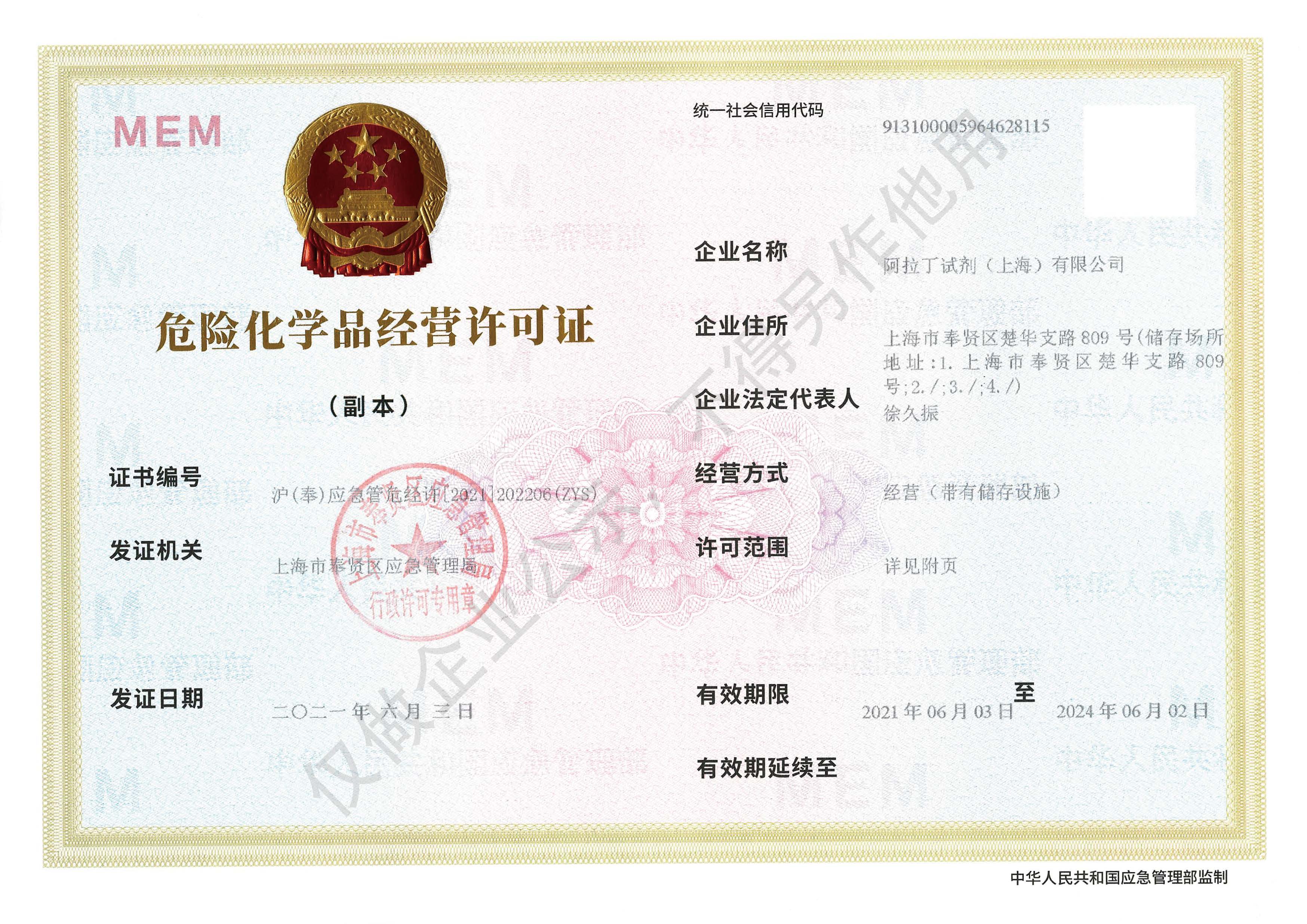 危险品化学品经营许可证(带存储)